
Minor Surgery Imaging Selector
Choose your surgical scenario to get recommended imaging modality.
When a surgeon tackles a minor procedure-think skin lesion removal, carpal tunnel release, or a small orthopedic fixation-the chance to picture the target before cutting can be a game‑changer. That’s where diagnostic imaging steps in, turning a blind guess into a precise plan and often sparing patients extra trips, complications, or surprise findings.
Key Takeaways
- Imaging sharpens surgical planning, reduces operative time, and boosts safety for minor cases.
- Ultrasound, X‑ray, CT and MRI each bring unique strengths; pick the one that matches the tissue, size and urgency.
- Integrating imaging into the workflow means pre‑op scans, intra‑op guidance and post‑op checks all work together.
- Avoid common pitfalls like over‑reliance on a single view or ignoring radiation dose limits.
- Emerging portable devices and AI‑driven interpretation are reshaping how quickly surgeons can see inside the body.
What is Diagnostic Imaging and Why It Matters for Minor Surgery
Diagnostic imaging is a set of techniques that create visual representations of internal anatomy, helping clinicians identify, localize and evaluate pathology without making an incision. While big‑ticket surgeries like organ transplants have long relied on imaging, its role in minor operations is often underestimated. A clear picture can reveal hidden cysts, calcifications or vessel anomalies that would otherwise surprise the surgeon mid‑procedure.
Beyond discovery, imaging supports three practical goals:
- Accurate targeting: Pinpoint the exact spot to cut or inject.
- Risk mitigation: Spot nearby nerves, arteries or tendons that need protection.
- Outcome verification: Confirm that the lesion is fully removed or the fixation is stable.
Choosing the Right Modality: A Quick Comparison
Not every scan fits every job. Below is a side‑by‑side look at the most common tools used in day‑to‑day minor surgery settings.
| Modality | Resolution (detail) | Radiation | Typical Cost (AU$) | Best For |
|---|---|---|---|---|
| Ultrasound | High (soft‑tissue) | None | 150‑300 | Superficial lesions, tendon or nerve mapping |
| X‑ray | Moderate (bone) | Low‑moderate | 80‑150 | Fracture confirmation, foreign body localization |
| CT Scan | Very high (cross‑sectional) | Moderate‑high | 400‑800 | Complex anatomy, deep‑seated cysts, pre‑op planning for bone work |
| MRI | Excellent (soft‑tissue, cartilage) | None | 600‑1200 | Ligament injuries, cartilage defects, soft‑tissue tumors |
When you’re dealing with a small skin nodule, the cheap, radiation‑free ultrasound will usually beat a costly MRI. If the case involves a tiny bone fragment, a quick X‑ray gets the job done. For anything deeper or more complex, a low‑dose CT or targeted MRI may be warranted.
Integrating Imaging into the Surgical Workflow
Seeing the problem before you cut is just the first step. The real power comes from weaving imaging through every phase of the operation.
1. Pre‑operative Planning
After the initial clinical exam, the surgeon orders the appropriate scan. The radiologist’s report should answer three questions:
- Where exactly is the lesion?
- What structures surround it?
- Are there any hidden risks (e.g., calcified vessels) that could change the approach?
Armed with these answers, the surgeon can mark the skin, choose the incision size, and decide whether a local anesthetic is sufficient or if a nerve block is safer.
2. Intra‑operative Guidance
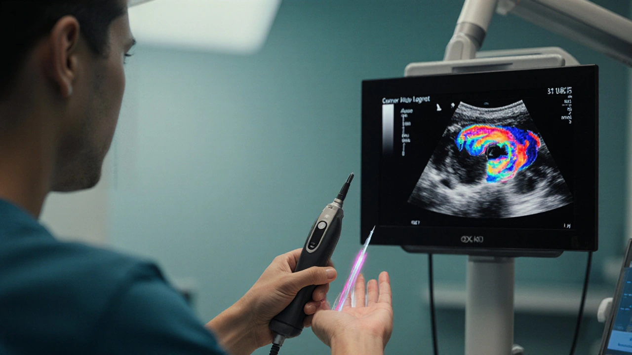
Some minor procedures happen in the clinic room with a handheld portable ultrasound probe. Real‑time images let the clinician watch the needle glide into a cyst or see a tendon slip away from a scalpel.
For slightly deeper work-like a percutaneous pin placement-fluoroscopy offers live X‑ray feedback. The surgeon can confirm pin trajectory without guessing, slashing both radiation exposure and the number of passes.
3. Post‑operative Verification
Even after the wound is closed, a quick scan can catch complications early. A bedside ultrasound can spot a developing hematoma, while a follow‑up X‑ray confirms that a tiny screw stayed where it should.
Documenting this final image also builds a clear record for future care-useful if the patient needs a repeat procedure down the road.
Pitfalls to Watch Out For
Even the best technology can backfire if you’re not careful. Here are the most common snags and how to dodge them.
- Over‑imaging: Ordering a CT for a superficial lipoma adds unnecessary radiation and cost. Stick to the lowest‑risk tool that answers the clinical question.
- Misinterpretation: Not all radiologists are familiar with the nuances of minor surgery. A brief discussion between surgeon and radiologist can clear up ambiguous findings.
- Equipment mismatch: Using a high‑frequency ultrasound for deep‑seated lesions yields blurry pictures. Choose the probe frequency that matches depth.
- Ignoring patient factors: Pregnant patients need radiation‑free options-ultrasound or MRI-unless absolutely unavoidable.
Future Trends: AI, Portability, and Point‑of‑Care Imaging
Technology isn’t standing still. Two trends are already reshaping how surgeons use imaging for minor procedures.
AI‑Assisted Interpretation
Machine‑learning algorithms can flag suspicious areas in seconds, letting the surgeon focus on the relevant slice. In a busy clinic, an AI‑powered ultrasound app can alert you to a hidden nerve bundle you might otherwise miss.
Handheld, Battery‑Powered Scanners
New handheld MRI units, while still niche, are beginning to appear in major hospitals. For minor orthopedic work, a portable low‑field MRI can give you cartilage detail without moving the patient to a full‑size scanner.
Integrated Surgical Platforms
Some modern operating tables now come with built‑in imaging arms that swivel into position, merging the pre‑op image with the live view. This reduces set‑up time and keeps the sterile field intact.
Putting It All Together: A Checklist for the Busy Surgeon
Before you walk into the procedure room, run through this quick list.
- Define the clinical question: What do I need to see?
- Select the least invasive, most informative modality.
- Confirm the radiology report addresses location, surrounding anatomy, and risks.
- Prepare any intra‑op imaging equipment (probe, fluoroscopy, etc.) and test it.
- Mark the skin based on imaging landmarks.
- Use real‑time imaging during the key steps of the procedure.
- Capture a post‑op image to verify success.
- Document findings, images, and any deviations from the plan.
Following this flow not only makes the surgery smoother-it also gives patients confidence that every angle was considered.

Frequently Asked Questions
Do I really need imaging for a simple mole removal?
Usually not. Most dermatologists rely on visual inspection and dermatoscopy. Imaging only becomes useful if the lesion is suspicious for deeper invasion or if you suspect involvement of underlying structures.
How much radiation does a typical hand X‑ray deliver?
A standard hand X‑ray imparts roughly 0.001 mSv-about the same as a few hours of natural background radiation. It’s considered safe for most adults, but we still avoid repeat exposures when possible.
Can I use ultrasound to guide carpal tunnel release?
Yes. Real‑time ultrasound lets you see the median nerve, flexor tendons and the transverse carpal ligament, helping you avoid nerve injury and keep the incision minimal.
What’s the biggest advantage of CT over plain X‑ray for minor orthopedic work?
CT provides cross‑sectional detail, letting you view the exact orientation of a fracture fragment or a hidden cyst that a single X‑ray plane might miss.
Is AI reliable enough to trust its suggestions during surgery?
AI is great as a second pair of eyes-highlighting patterns, measuring dimensions, and flagging anomalies. However, the final decision should always rest with the surgeon, who considers the whole clinical picture.


Brian Skehan
Everyone knows the real reason imaging is getting so pushy in minor surgery-it's a data‑harvesting goldmine for the shadow agencies that think they can track every incision you make. They slip tiny RFID‑like tags into the ultrasound gel and claim it's "contrast." It’s not about safety; it’s about surveillance under the guise of patient care. If you think the surgeon cares, think again.
Andrew J. Zak
Imaging can be helpful but let’s keep the focus on patient outcomes
Maureen Crandall
Honestly the whole checklist feels like a bureaucratic nightmare we dont need it is just another way to complicate a simple mole removal
Michelle Pellin
Dearest colleagues, the symphony of light that emanates from a handheld ultrasound is nothing short of poetic, a ballet of echoes that pirouette across dermal layers, revealing secrets once hidden beneath the epidermis. When we wield such a device in the delicate theatre of minor surgery, we are not merely cutting tissue; we are orchestrating a masterpiece where precision and compassion entwine. Let us therefore celebrate the convergence of technology and art, for therein lies the true renaissance of surgical practice.
Keiber Marquez
We dont need fancy MRI for a tiny skin bump its overkill and waste of tax money the US should stick to basic Xray for real problems
Lily Saeli
In the grand tapestry of human health, the pursuit of ever‑more imaging tools threatens to transform our bodies into mere data points, stripped of dignity and sanctity. It is a moral imperative to ask whether the relentless quest for visual clarity supersedes the humble wisdom of clinical judgement, lest we become servants to the microscope rather than its masters. Let us temper our curiosity with compassion, remembering that every scan echoes a deeper responsibility toward the patient’s wholeness.
Joshua Brown
The integration of diagnostic imaging into minor surgery begins with a clear clinical question, which serves as the compass guiding modality selection, ensuring that the chosen tool answers precisely what the surgeon needs to know. First, assess the anatomical depth of the lesion; superficial structures such as skin lesions or tendon paths are exquisitely visualized with high‑frequency ultrasound, a modality that offers real‑time feedback without ionizing radiation, thereby preserving patient safety. Second, consider bone involvement; conventional radiography (X‑ray) provides rapid, low‑dose assessment of fractures or foreign bodies, delivering sufficient detail for most outpatient scenarios. Third, when confronted with complex, deep‑seated cysts or intricate bony architecture, computed tomography (CT) delivers cross‑sectional detail, albeit at the cost of higher radiation exposure, which must be judiciously weighed against diagnostic benefit. Fourth, magnetic resonance imaging (MRI) excels in soft‑tissue contrast, cartilage assessment, and ligamentous injuries, presenting an unparalleled view of the internal landscape while remaining radiation‑free, though accessibility and cost may limit its routine use. In practice, the surgeon should collaborate closely with the radiologist to refine the scan protocol, tailoring slice thickness, plane orientation, and contrast administration to the procedural goals. Pre‑operative imaging findings should be transcribed onto the operative field, either by skin marking or digital overlay, allowing the surgeon to navigate with confidence. Intra‑operatively, portable ultrasound probes can be sterilized and positioned directly over the surgical site, granting live visualization of needle trajectories, tissue planes, and vascular structures, thereby reducing the risk of iatrogenic injury. Fluoroscopic guidance, when employed, demands strict adherence to ALARA principles, using pulsed dose settings and shielding to minimize exposure for both patient and staff. Post‑operative verification, such as a bedside ultrasound for hematoma detection or a follow‑up X‑ray for hardware placement, serves as a quality assurance measure, confirming procedural success before the patient leaves the clinic. Documentation of each imaging step, including parameters, findings, and any deviations from the original plan, creates an audit trail that supports continuous improvement and medicolegal protection. Moreover, emerging artificial intelligence algorithms can flag subtle anomalies in real time, acting as a second set of eyes that enhances diagnostic confidence without replacing clinical judgment. Portable low‑field MRI systems are beginning to enter ambulatory settings, offering cartilage detail that was once confined to major academic centers, expanding the horizon of what is possible in a minor procedure suite. Finally, a concise checklist-defining the question, selecting the modality, confirming report completeness, preparing equipment, marking landmarks, employing real‑time guidance, capturing post‑op images, and recording outcomes-ensures a systematic, patient‑centered approach that maximizes safety and efficacy. By adhering to these principles, surgeons can transform minor interventions from blind guesses into precisely choreographed performances, ultimately improving patient satisfaction and clinical results.
andrew bigdick
Hey folks, love the deep dive-if you're ever unsure whether to grab an ultrasound or just go with a quick X‑ray, think about how deep the problem sits and how much radiation you want to give the patient.
Shelby Wright
While the checklist you laid out is thorough, it's also a textbook example of how we can drown simple procedures in bureaucratic excess-sometimes the surgeon's intuition should trump a 15‑step protocol, especially when time is of the essence.
Ellen Laird
One must question whether the proliferation of high‑tech imaging in minor surgeries is a true advancement or merely a fashionable indulgence for those who cant resist the lure of shiny equipment.
rafaat pronoy
Cool stuff overall 😊 just remember the patient’s comfort is key, and a quick scan that’s easy on them wins every time.