Preoperative Imaging: Why It Matters for Every Surgery
When working with preoperative imaging, the set of diagnostic scans performed before a surgical procedure to map anatomy, identify pathology, and plan the operative approach. Also known as pre‑op imaging, it provides the roadmap surgeons rely on. Commonly, a CT scan, a cross‑sectional X‑ray that visualizes bone and soft tissue in high detail is ordered for orthopedic or trauma cases. MRI, magnetic resonance imaging that captures soft‑tissue contrast without ionizing radiation shines in neurologic and musculoskeletal assessments. When real‑time imaging is needed, an ultrasound, high‑frequency sound waves that produce live images of organs and vessels becomes the tool of choice. Finally, contrast agents, substances injected to enhance blood vessels and lesions on scans often boost the diagnostic power of CT and MRI, helping surgeons spot hidden issues before cutting.
Preoperative imaging isn’t just about taking pictures; it directly influences surgical strategy. The detailed anatomy revealed by a CT scan can dictate incision location, screw length, or implant size. MRI findings may alter the decision to use minimally invasive techniques versus open approaches, especially when nerves or tumor margins are involved. Ultrasound guidance can prevent accidental organ injury during laparoscopic entry. In each case, the imaging data become a shared language between radiologists, surgeons, and anesthesiologists, ensuring everyone is on the same page before entering the operating room.
Selecting the right modality starts with the clinical question. If bone detail is paramount—think spinal fusion or fracture repair—a CT scan usually wins. When soft‑tissue contrast matters, such as evaluating a brain tumor or ligament tear, MRI provides the clarity needed. For bedside assessments or fetal surgery, portable ultrasound offers speed and safety. Sometimes, a combination is required; a CT angiogram with contrast might map vascular anatomy before a cardiac bypass, while an MRI with gadolinium highlights inflammatory tissue before joint replacement. Understanding these nuances helps clinicians avoid unnecessary tests and keeps patients from extra radiation exposure.
Contrast agents add another layer of decision‑making. Iodinated contrast for CT can highlight blood vessels, tumors, and infections, but it demands kidney‑function checks to prevent nephrotoxicity. Gadolinium‑based contrast for MRI enhances lesion detection but carries a small risk of deposition in the brain, especially with repeated use. Choosing the appropriate agent involves weighing diagnostic benefit against patient safety, reviewing allergy history, and coordinating with the pharmacy team. Proper timing of contrast administration and image acquisition maximizes image quality and reduces repeat scans.
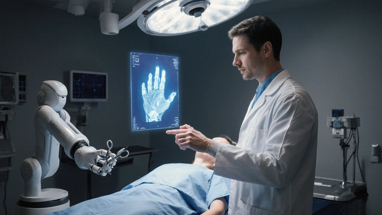
Interpreting preoperative images is a collaborative effort. Radiologists generate detailed reports, highlighting critical findings, measurement of lesions, and any incidental issues that could affect surgery. Surgeons review these reports alongside the images, often using 3‑D reconstructions or virtual reality tools to visualize complex anatomy. Anesthesiologists look for airway challenges or vascular access concerns noted on scans. This multidisciplinary dialogue transforms raw data into actionable plans, reduces intra‑operative surprises, and improves postoperative outcomes.
Below you’ll find a curated collection of articles that dive deeper into each imaging modality, contrast considerations, and real‑world case studies. Whether you’re a surgeon fine‑tuning your pre‑op checklist, a radiologist seeking optimization tips, or a patient curious about the scans you’ll undergo, the resources ahead will give you practical insights and actionable guidance.